
In Depth: Effects of CO-CR Discrepancy in Daily Orthodontic Treatment in Planning
Introduction:
Within the dental profession there is a general consensus that treatments that modify the occlusion should be planned in centric relation (CR). Centric relation is defined as musculoskeletal stable position, with the condyles forward, as far upward, centred transversely and with the articular disc interposed properly.
Centric relation (CR) is a musculoskeletal position, anatomically determined, repeatable and reproducible. Treating our patients in this position is one of our biggest challenges, both in orthodontics and prosthodontics. This ideal goal will provide long-term stability, improved functionality and aesthetics, healthy musculature and stable temporomandibular joints.
On the other hand, centric occlusion (CO) or maximum intercuspation is a dentally determined position. Tooth morphology and position are the primary influences determining the mandibular position and movements. The condylar position is strongly determined by the dental contacts and intercuspation.
These two reference positions of the mandible do not usually coincide in natural dentition with a ‘normal’ minor, acceptable slide. If the dental occlusion is not stable or if there are significant premature contacts the mandible will have an abnormal CO-CR slide, leading to secondary effects such as abrasion, deviation of mandible, TMJ signs and symptoms and skeletal asymmetry.
A destabilised occlusion essentially causes a repositioning of the mandibular condyles in the glenoid fossa. This can be uncomfortable for patients in many ways.
Historically, the most common way to classify the ideal static state of the dentition was Angle’s molar relationship. This classification, along with clinical findings, was generally enough for orthodontists to make a diagnosis and decide on a treatment plan.
However, the gold standard is to diagnose our cases in a CR analysed occlusion (on articulator mounted models). The discrepancy between CO and CR has been noted to be present in most of the cases. We may find an increased overjet, reduced overbite and Angle’s molar relationship modification - all of these possibly leading to a significant change in diagnostic conclusions and therefore the treatment plan and subsequent treatment mechanics.

SEVERAL METHODS ARE GIVEN IN ORDER TO EVALUATE THE DISCREPANCY BETWEEN CO AND CR:
• Direct visual evaluation in the oral cavity
• Articulated casts with CR bite registration
• Radiological and 3D imaging
• MCD (measures condyle displacement)/CPI (condyle position indicator) tool
• Modjaw System.
THE CLINICAL RECORDS TAKEN FOR EACH ORTHODONTIC PATIENT WOULD IDEALLY CONSIST OF:
1. Written dental and medical history.
2. Comprehensive clinical examination (including facial structures, masticatory muscles, static and dynamic occlusion and TMJ assessment).
3. Study cast models (and/or digital impressions) with 2 types of bite registrations :
- CO bite registrations in pink wax.
- CR bite registrations in blue Almore wax( centric “ du jour”).
4. Facebow registration of the terminal estimated hinge axis position.
5. Graphic three-dimensional position of the mandibular condyles using the MCD/CPI tool or the Modjaw System.
6. Facial and Dental Photographs.
7. Orthopantomography and lateral Cephalometric X-rays
8. Cone Beam CT or MRI scans in certain specific cases
THE FOLLOWING ORTHODONTIC TREATMENT SEQUENCES CAN BE INFLUENCED BY THE PRESENCE OF A CO-CR DISCREPANCY
1. DIAGNOSIS PHASE:
• the magnitude of the horizontal, vertical and transversal discrepancy to be corrected
• the lateral cephalometric radiograph analysis changes + skeletal changes in sagittal
• the mandibular autorotation anticipated throughout treatment
• True / psedo-mandibular asymmetry
• the possible presence of TMD conditions and the patients susceptibility to TMD
2. TREATMENT PLANNING PHASE:
• extraction vs. non-extraction treatment
• orthodontic treatment only vs. orthodontic-surgical treatment
• the treatment mechanics (given especially by the diagnosis and the anchorage needs)
• the final occlusal finishing of the case (the position of the dental arches in a three-dimensional approach)
3. REQUIREMENTS FOR A MULTIDISCIPLINARY APPROACH TO THE CASE
4. THE POSSIBLE RELAPSE DEGREE OF THE CASE
Case Example 1:
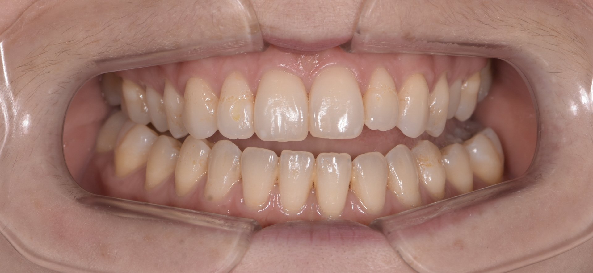
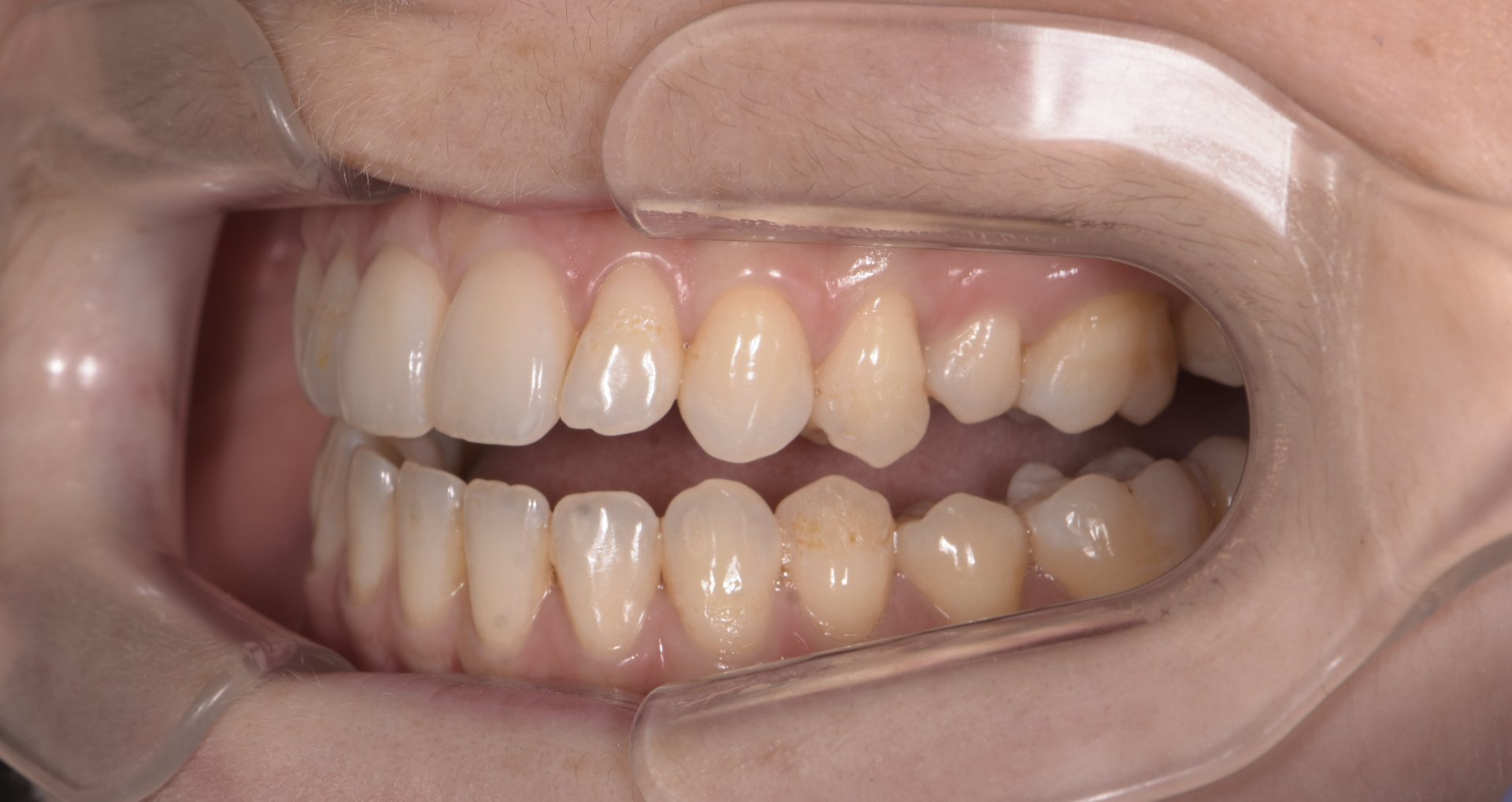
46 year old female who had orthodontics as a teenager presented with TMJ pain and headaches in the morning.
No long term retention, upper wisdom teeth present.
CR records showed only one contact on the upper right second molar and lower right second molar.
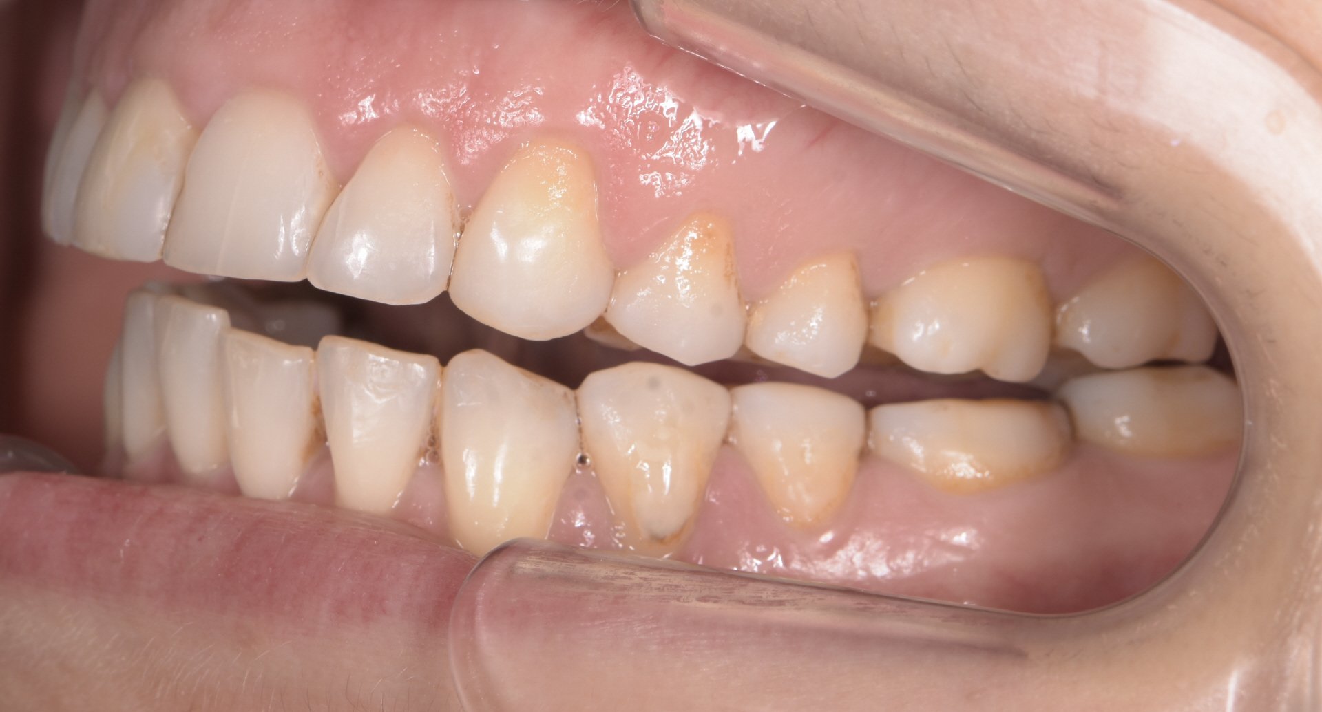
Level of Case Difficulty: MAXIMUM
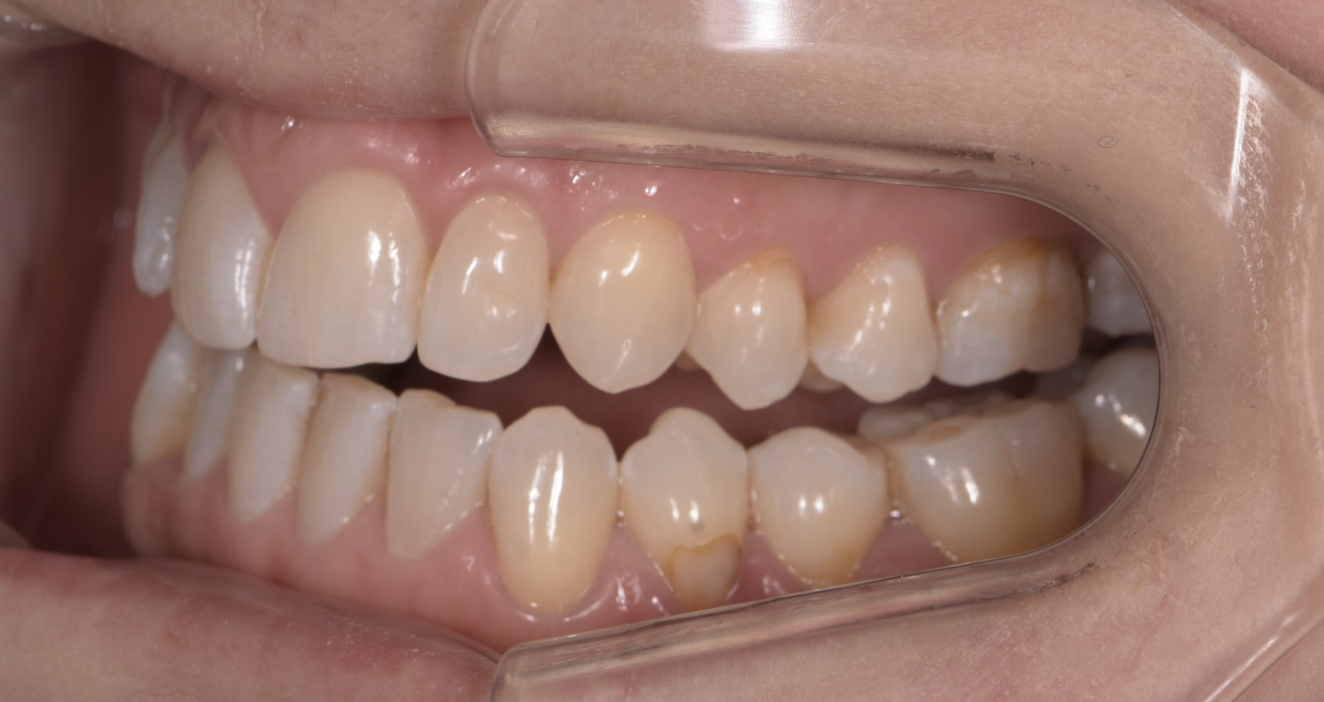
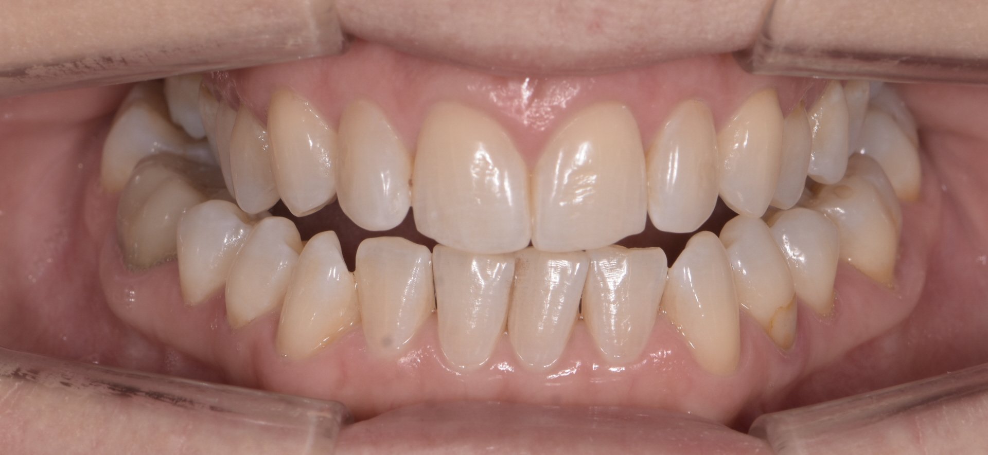
Case Example 2:
35 year old female. Previous orthodontics with lingual braces.
Presenting complaint : unable to bite in stable position and teeth do not meet.
CR records and articulated casts mounted in CR showed only one contact on the upper left second molar and lower left second molar.
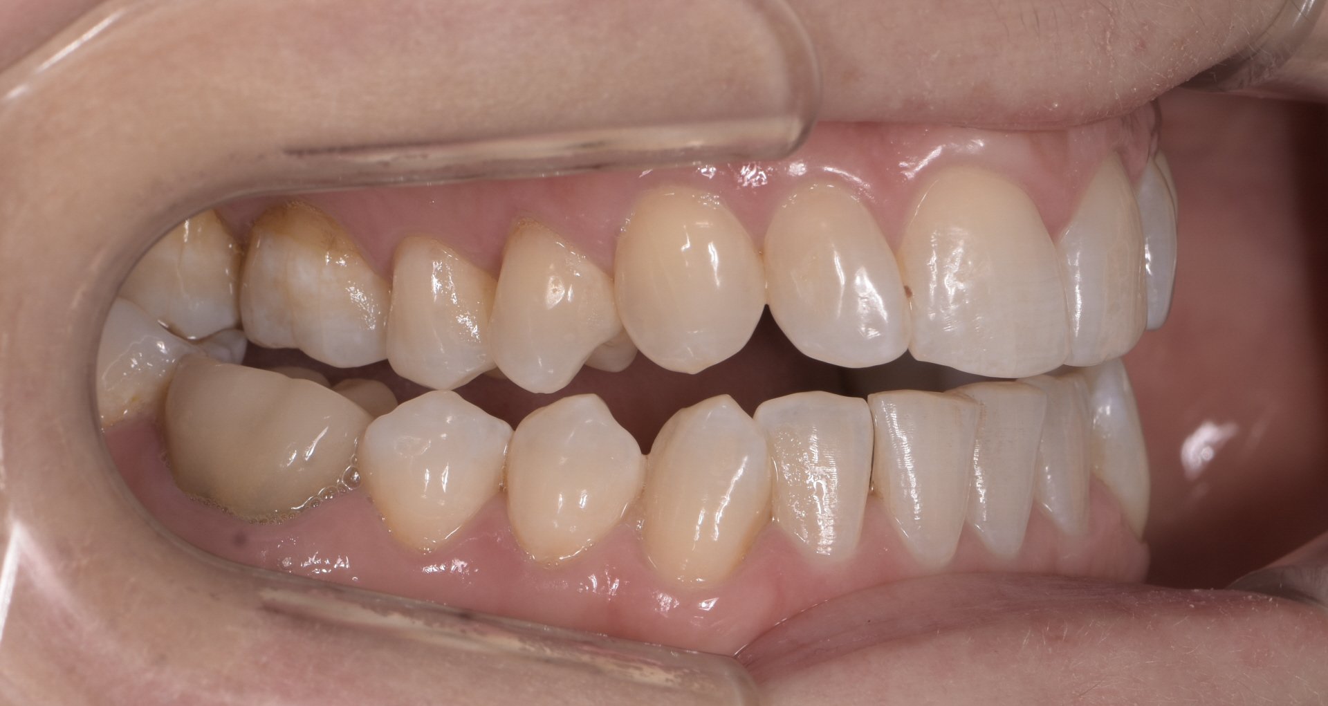
Level of case difficulty: Maximum
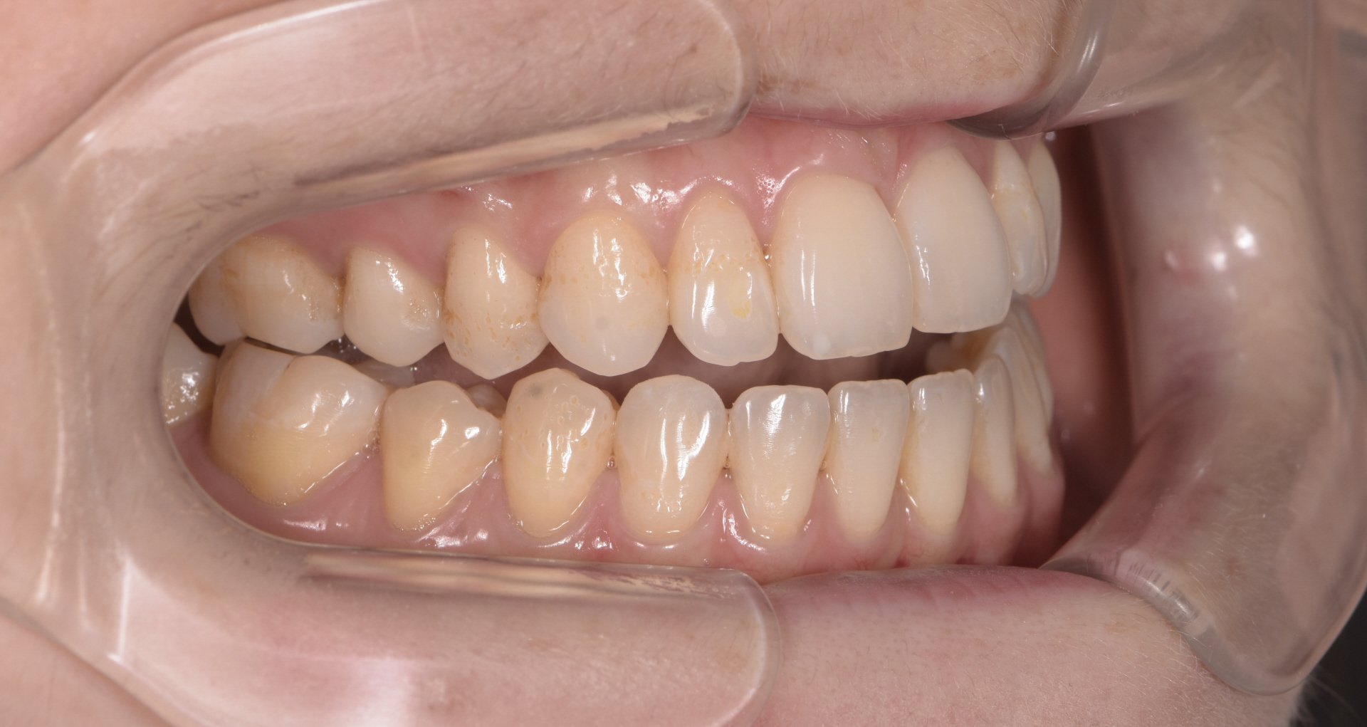
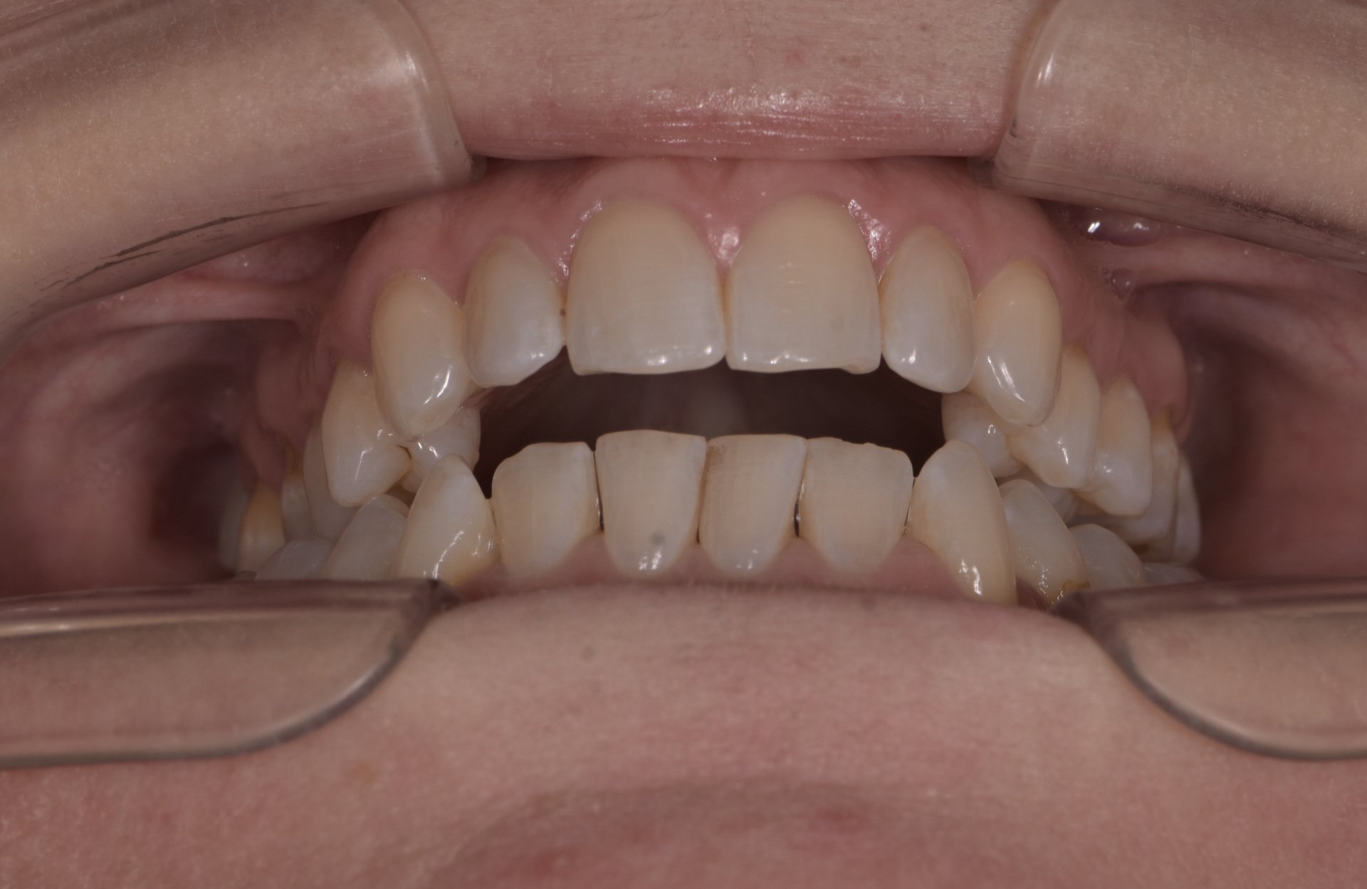
Case Example 3:
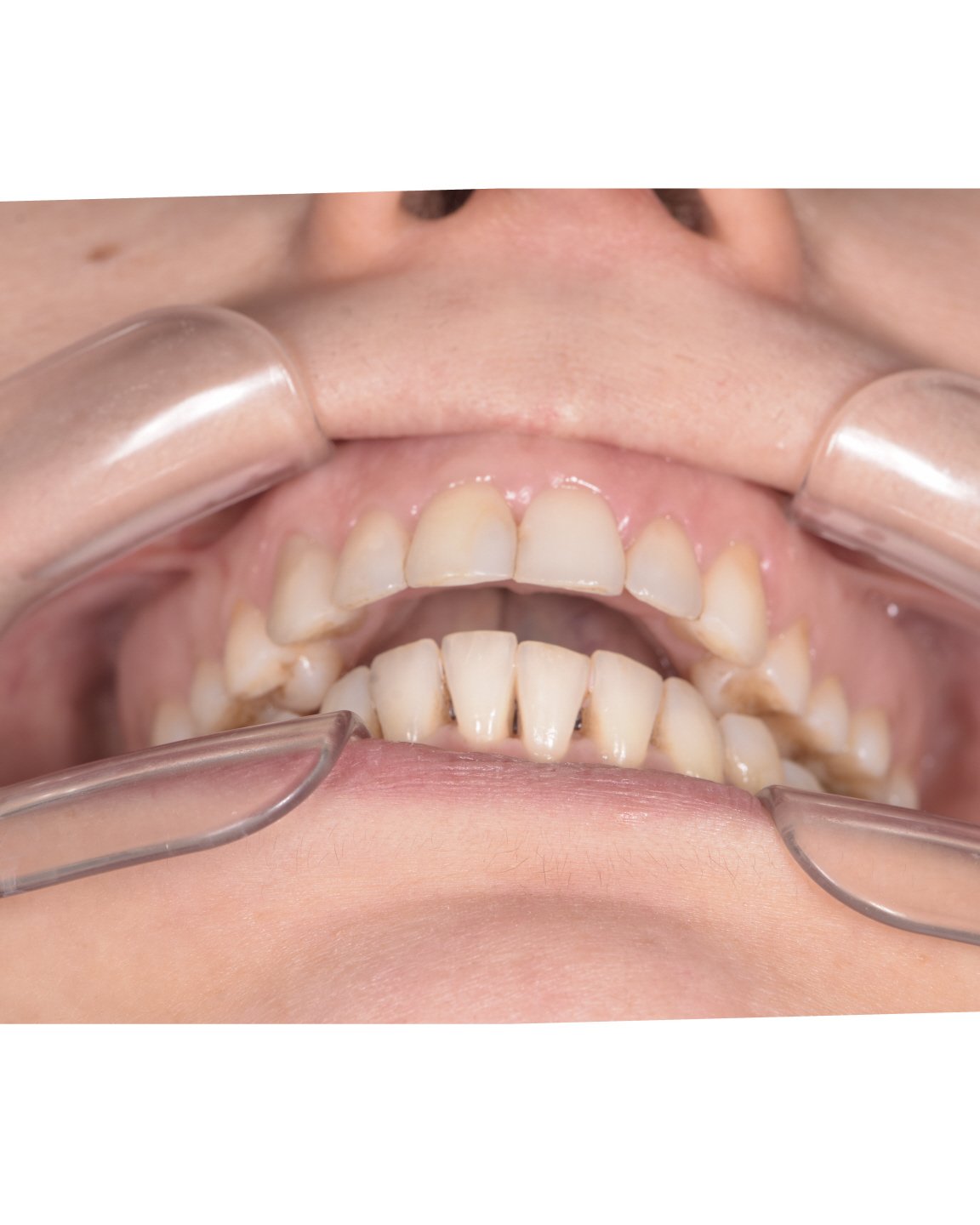
25 year old female. Previous orthodontics as a teenager.
Came to check her lower fixed retainer which was poking her.
Bite has been monitored by GDP.
CR records showed only contact on the upper right first molar and lower right first molar.